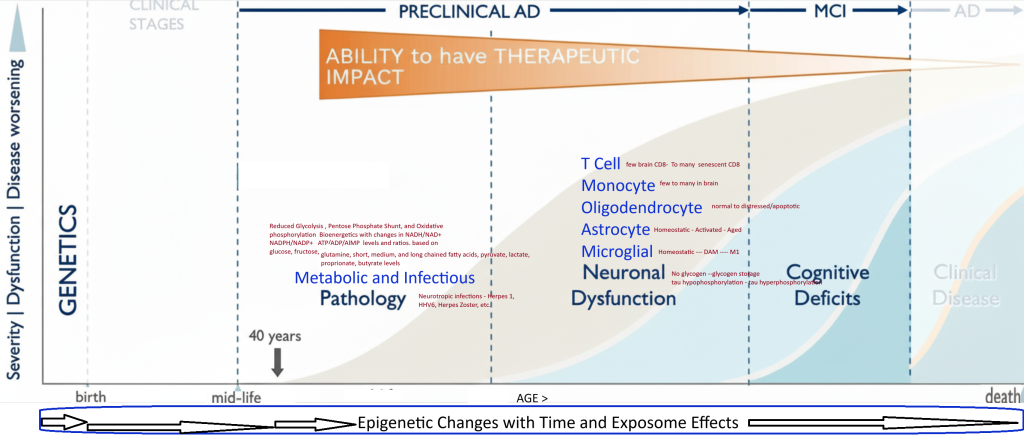
MCI and All Dementias : EOAD and LOAD
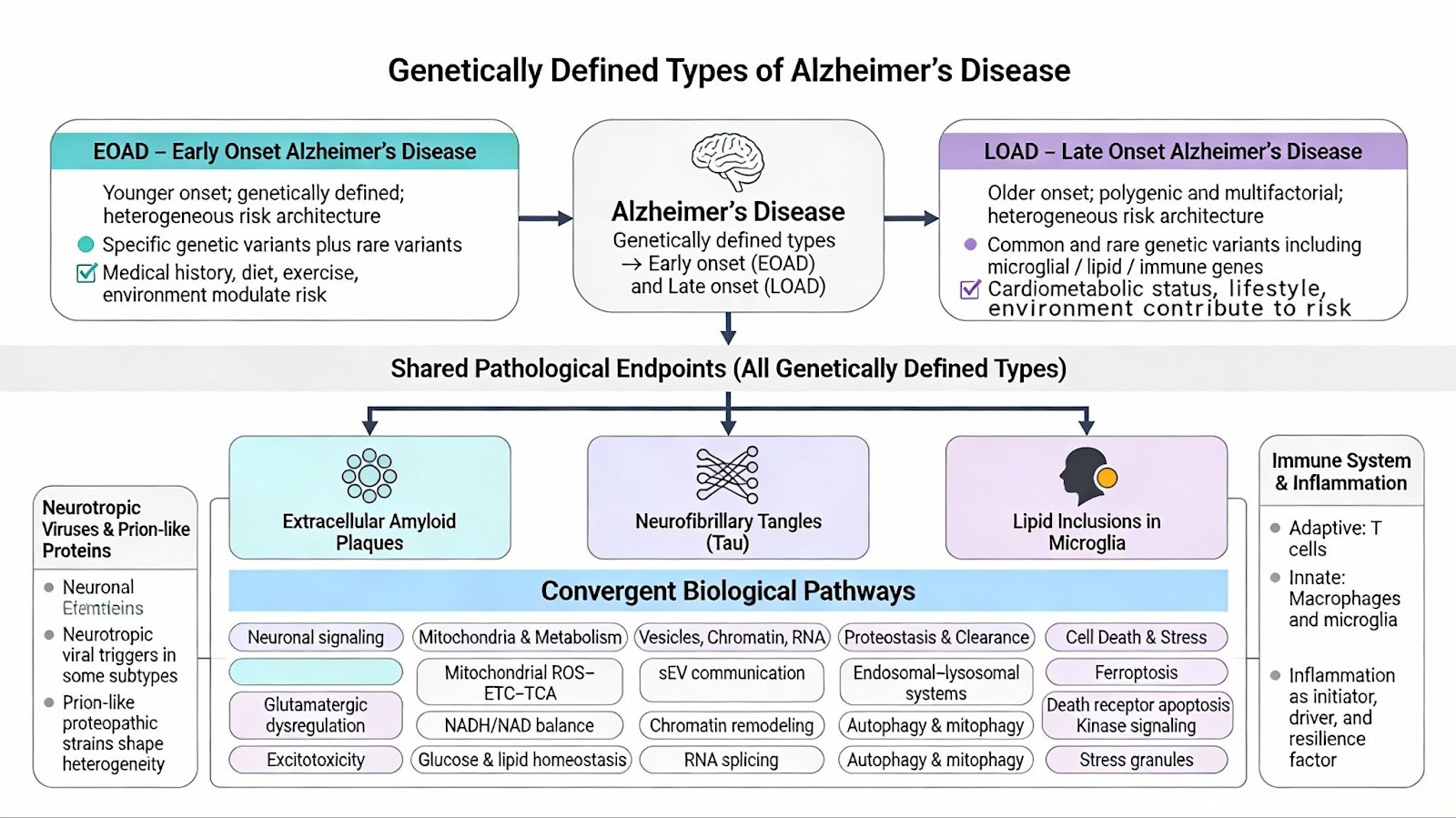
Genetically defined types of Alzheimer’s disease can be subtyped into either early or late onset. All of the reported genetically defined types share the common endpoint pathology of Neurofibrillary Tangles, Lipid inclusions in Microglia, and Extracellular Amyloid Plaques. Both LOAD and EOAD are heterogeneous diseases that are caused by specific genetic and exposome factors (, medical history, diet, exercise, and environmental factors). EOAD has about 90 % sporadic component and 10% familial, while LOAD has a 70% Polygenic component and a 30 % Sporadic subtype. Multiple frameworks to describe AD have been proposed including, Complex Biological System Models with AD subtypes , AD as an offshoot of the Biology of Aging, The Multiple hit theory of Genetics, Oxidative stress, chronic inflammation, and disrupted electron energy homeostasis
LOAD – Late Onset Alzheimer’s Disease
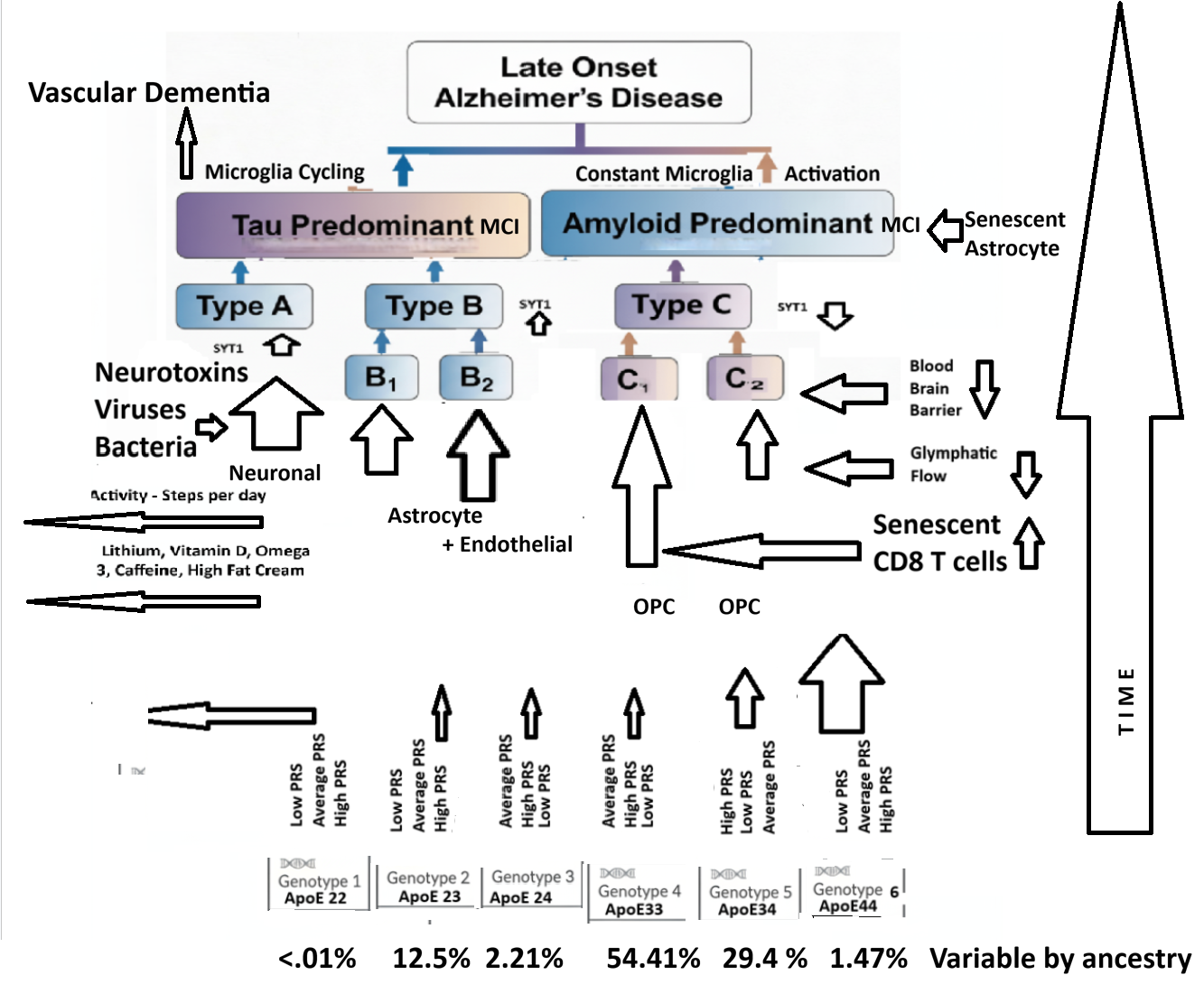
ApoE genotype stratified patients differentially express proteomes leading to different LOAD Neuropathology Types depending on their Exposome and Polygenic Risk Factors. Exposome factors include HSV1, Lithium in diet, Infant exposure to added sugar, Mediterranean diet, Coffee consumption, High Fat Cream Consumption, Microbial Ferulic Acid Production in the Intestine, Physical Activity Level, Medications including levetiracetam- Keppra, Education Level, Social Experiences, Brain Exercises, Periodontitis, and/or other Risk Factors .
Amyloid Predominant C1/C2
| Stage | Event | Key Consequences |
| 0 | Genetics for Amyloid predominant – = ApoE4, Amyloid gene duplication/Down’s Syndrome, Presenilin 1,2 . | Dysfunctional amyloid 42 production |
| 1 | Innate Metabolic Cascades and Mitochondrial Dysfunction | Reduced ATP/NADH Energy Production |
| 2 | Aging- decreased DNA methylation —> Line-1 /SVA activation | Retrotransposon derepression, cGAS-STING overactivation– Senescence |
| 3 | Pathogenic App specific CD8 T cell and Monocyte-Microglial brain infiltration | Interferon gaama, perforin, SASP factors, Chronic Neuroinflammation |
| 4 | Neuronal/Astrocyte/OPC-OL/Microglial Cell Death and Neuronal Network Failure | Karyoptosis, Apoptosis, synaptic loss, Progressive Neurodgeneration |
33% C1 – OPC with High Microglial Contribution – Involving pathogenic CD8 T Cells and APoE4 influenced
C2 – OPC with Low Microglial Contribution – A primary Oliodendrocytopathy – not involving CD8 T cells.
Tau Predominant Types A, B1, B2
| Stage | Event | Consequence |
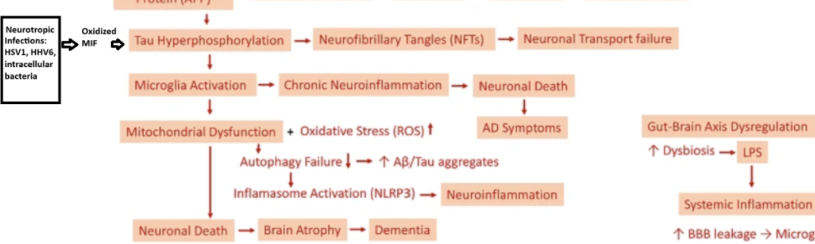
| 0 | Birth – HLA-DR risk genes for neurotropic infections | Tau and Amyloid production |
| 1 | HSV1 in hippocampal glutamatergic neurons or other brain cell infection | Chronic neuronal/astrocyte/microglial/OPC stress and oxidative environment |
| 2 | Oxidized alpha MIF Signaling | Tau phosphorylation and oligomer formation |
| 3 | Tau oligomers activation of Transposons | cGAS-STING activation by LINE-1 elements and senescence induction |
| 4 | anti neurotropic CD8 T cell infiltration into brain | Either protective or destructive T cells impact on the brain |
| 5 | Cell Death and Network Failure | Apoptosis, necroptosis, synaptic loss, progressive neurdegeneration |
Tau Predominant seen in microglial methylome data, brain transcriptome signatures, and Tau PET Scan analysis
33% A Neuronal dysfunction predominant – Most likely connected to Infection Related Sporadic AD of both Late and Early versions. May be related to Exitotoxicity – Mitochondrial dysfunction and Apoptosis pathway contributions.
33% B1 Astrocyte Predominant – Blood Brain Barrier and Glymphatic Flow – Amyloid Clearance connected and
B2 Endothelial then Astrocyte Predominant – CardioVascular Disease and Vascular Dementia connected
Amyloid Predominant seen in microglial methylome data and brain transcriptome signatures

Pathological mechanisms and treatment progression of Alzheimer’s disease | European Journ
All Pathways